1.1 Womb as environment
It has become very evident that the placenta is a multifunctional organ that integrates transport, endocrine, immune, and epigenetic processes, to actively regulate fetal growth and development, acting more of a control center, than previously thought, as a passive transporter like a bridge (Chen et al., 2024; Danhausen & King, 2024).
The placenta
The placenta develops within the uterus of a pregnant person. Very early in human prenatal development, the zygote develops into a blastocyst which has an outer layer of cells, called trophoblasts, as well as an inner cell mass. This inner cell mass is what will develop into the embryo. The trophoblast will attach to the rich endometrium of the uterus and eventually form the placenta. The maternal-placenta-embryo circulation is in place by the end of the fourth week, and the placental structure is complete by the twelfth week, allowing for the exchange of gas, nutrients and waste between the embryo and the pregnant individual. As the pregnancy continues, the placenta continues to grow and cover about half of the uterine surface by 5 months, increasing the functional surface area (Keenan-Lindsay, 2022; Moore et al., 2020).
In the video below, Dr. Alan Bocking, professor in the Department of Obstetrics and Gynaecology at the University of Toronto, explains more about the main functions of the placenta.
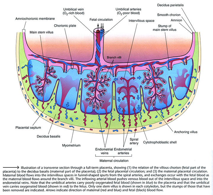
Visualize from the diagram below how blood flows through a fully developed placenta. Some of the main processes which occur through this system involve transfer of oxygen and nutrients from the gestational parent to the fetus and waste and carbon dioxide from the fetus to the gestational parent.
Bocking describes the placenta as an “incredibly impressive endocrine organ”. Play the “Build a Baby” game to learn about the four main hormones produced by the placenta and how they work to support pregnancy.
As an essential organ which only develops during pregnancy, the placenta plays a crucial role in the quality of the intrauterine environment experienced by the developing fetus. The placenta is capable of adapting its function to a variety of conditions as it integrates chemical messages from the pregnant individual and the fetus. For instance, the placenta will signal the maternal system when the fetus needs certain nutrients or hormones (Danhausen & King, 2024).
How well the placenta is able to develop and function has important implications for fetal growth and development. Listen as Dr. Stephen Lye explains more about these implications.
Moffett-King, A. (2002), Natural killer cells and pregnancy, Nature Reviews Immunology, 2, 656-663. doi: 10.1038/nri886 Retrieved from http://www.cvs.ed.ac.uk/sites/default/files/A%20Moffett-King_0.pdf
Copyright 2002.
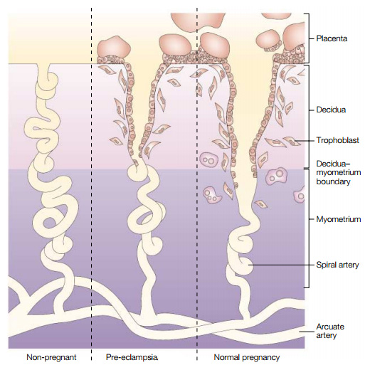
According to Moore et al. (2020), 80 – 100 spiral arteries are found within the decidua basalis (a maternal part of the placenta seen in the first diagram on the page). Blood flows from these spiral arteries into the intervillous space. Exchange of products with the fetus occurs as blood from the pregnant person flows more slowly around the branch villi (p. 69). In the second diagram on the page, note the variance in quality of spiral artery invasion from trophoblast cells that may occur during pregnancy. With preeclampsia, the condition mentioned in the preceding video, this invasion occurs at a superficial level. In a normal pregnancy, the trophoblast cells invade into the myometrial, not just the decidual portion of these blood vessels and transform the spiral arteries into more open vessels. The structural changes in this healthier invasion accommodate a significant increase in uterine blood flow (optimizing the volume of blood flowing through this location in the fetal supply line) Danhausen & King, 2024, p. 753).
If you are interested in further information on preeclampsia, read FAQs on the Preeclampsia Foundation website.
Uteroplacental blood flow is essential for delivering oxygen and nutrients to the fetus, and it depends on healthy maternal blood vessels and proper placental development. Conditions such as pre-eclampsia(hypertension), diabetes, anemia, infection, poor nutrition, stress, as well as behaviors (drug use/smoking) can reduce blood flow by increasing vascular resistance, damaging blood vessels, or lowering oxygen-carrying capacity. When placental blood flow is impaired, less oxygen and nutrients reach the fetus, which can lead to complications, such as fetal growth restriction and preterm birth impacting growth and development of the fetus with potential long-term implications (Chappell et al.,2023; Li et al., 2018; Hu & Zhang, 2021).
Listen to the next video as Dr. Maggie Morris describes how smoking impacts placental function.
Just as concerning with pregnant individuals smoking and the effects on the placenta and fetus, nonsmoking pregnant individuals may still be exposed to tobacco smoke through household members, making them “passive smokers.” This exposure is associated with adverse outcomes including low birth weight, infant morbidity and mortality, and long-term developmental concerns, such attention, learning and behavioral issues in the offspring (Berk, 2022, p. 68).
Read more about how smoking in pregnancy can affect the gestational parent and fetus in the following synthesis of information on the Encyclopedia on Early Childhood Development website.
Stress in the womb
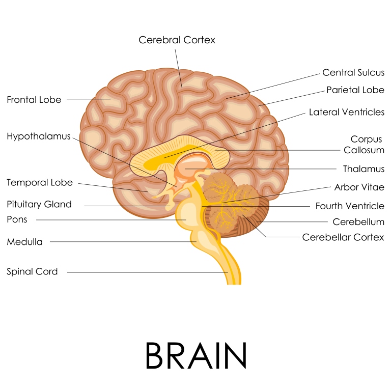
Prenatal stress is the physiological and psychological stress experienced during pregnancy, arising from factors such as anxiety, depression, trauma, socioeconomic challenges, or environmental exposures. These stressors, whether acute or chronic, can shape the environment in utero in a complex manner. The effects on the fetus are mediated through mechanisms including hormonal changes, epigenetic modifications, and alterations in placental function. Maternal stress has the potential to trigger a cascade of events within the body known as the hypothalamic-pituitary-adrenal (HPA) axis activation response increasing the production of stress hormones, in particular cortisol (Georgousopoulou et al., 2026; Jagtap et al., 2023). The body has an incredible response system to help it cope with stressors. One part of this system, as mentioned above, is the hypothalamic-pituitary-adrenal (HPA) axis. The hypothalamus, located in the forebrain, is situated just above the pituitary gland and helps the body maintain internal balance. It functions as a link between the nervous and endocrine systems. Watch the Endocrine system video from the MedlinePlus webpage Endocrine glands to see how the nervous and endocrine systems work together to maintain equilibrium within the body.
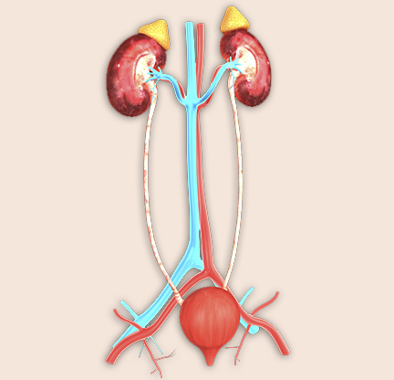
In the image on the right, note the adrenal glands, a set of triangular shaped glands that belong to the endocrine system, situated at the top of each kidney. Cortisol, a glucocorticoid hormone, is produced by the adrenal cortex (the outer segment of these glands) in response to either physical or psychological stress. Dr. Joanne Weinberg, professor emerita and distinguished scholar in the Faculty of Medicine at the University of British Columbia, explains how the HPA axis works both in response to stress and in situations of normal daily living.
Glucocorticoids are essential for life, influencing virtually every tissue and affecting a wide range of physiological functions from metabolism, blood pressure, the immune system, regulation of fluid and electrolyte homeostasis and increasing energy demands in response to stress” (Reynolds, 2013, p. 3).
Click through the next interactive activity to learn more about how the HPA axis works as a negative feedback system to help the body cope with more acute stressors.
According to Georgousopoulou et al. (2026) and McEwen (2017), the HPA axis provides protection to various body systems when activated. It has an impact on a variety of different regions in the brain that are responsible for emotional regulation, memory, and cognitive function. For example, in an acute threatening situation, memory may be improved from the release of cortisol, improving one’s ability to avoid similar dangerous situations in the future.
However, as discussed by Weinberg in the previous video, if a stressor were to become repeated or chronic, the prolonged activation of the HPA axis could result in more negative effects, such as illness related to suppression of the immune system. In the example of memory, which may be improved in an acute stressful situation, impairment might develop if the stress is chronic, increasing wear and tear on the brain and body systems resulting in altering one’s brain structure and function, as well as an increased risk to disease (McEwen, 2017; Roberts & Karatsoreos, 2021).
In the next video, Dr. Brandy Wicklow describes fetal development of the HPA axis, which will become the lifelong regulator of cortisol secretion.
Ross and Desai (2017) explain that for normal prenatal maturation and development of organs such as the kidneys, lungs and brain to occur, the fetus needs steroid hormones (e.g., the glucocorticoid hormone cortisol). They also point out that starting from midgestation, glucocorticoid receptors are expressed in most fetal tissues. The diagram of the brain presented earlier within the interactive activity shows the location of the hippocampus, an area known to have numerous glucocorticoid receptors. Learning and memory are important functions associated with the hippocampus (Ross & Desai, 2017, p. 93).
In the next video, Dr. Stephen Matthews explains more about maternal prenatal adversity, revealing that glucocorticoids are linked with programming of how the fetus functions after birth.
In a review article, Moisiadis and Matthews (2014a) describe how mounting scientific evidence supports the hypothesis that raised fetal cortisol levels associated with stress induced increases in maternal cortisol, can affect fetal neurological (brain) development as well as the function of the HPA axis after birth (pp. 397-398). They state conditions of maternal adversity such as depression, anxiety or undernutrition may result in increased levels of glucocorticoids within the mother and fetus (p.391). Additionally, they indicate physical stressors such as fetal hypoxia (lack of sufficient oxygen supply to its tissues) may also result in increased fetal glucocorticoid levels (p.391). Another originating cause of elevated fetal glucocorticoid levels internal to the maternal-fetal-placental unit they describe could occur when higher glucocorticoid levels in the maternal circulatory system are present and the placenta is not functioning adequately in its protective role (Chapman et al. as cited in Moisiadis & Matthews, 2014a, pp. 391-392).
Importantly, alterations in the set-point of the HPA axis during prenatal development from exposure to excessive glucocorticoids in utero may mold the development and function of the offspring HPA axis activity even into adulthood (Jagtap et al., 2023; ), which can have behavioural, cognitive and emotional consequences for the offspring (Georgousopoulou et al., 2026).
However, McGowan and Matthews (2018) point out the relationship between maternal prenatal adversity, fetal exposure to glucocorticoids, and subsequent effects on HPA axis function and reactivity to stress post-birth is complex. In their mini-review, they report factors such as the type and timing of prenatal adversity, offspring sex, context of the post-birth stressor activating the HPA axis as well as the age of the infant or child may all be contributing factors in how the HPA axis functions and how the infant or child responds to stress.
For example, in relation to offspring sex differences, research is demonstrating that male fetuses, generally, are more susceptible to adverse conditions in utero. Research indicates that exposure to elevated maternal cortisol is associated with changes in brain connectivity, larger amygdala (processing emotion, memory and behavior) volume, and increased stress reactivity in males. In contrast, female fetuses may demonstrate adaptive responses that help buffer against these effects, contributing to greater resilience (Georgousopoulou et al., 2026).
Also, additional research suggests that male fetuses may be more vulnerable to physical stress in the womb, while female fetuses may be more sensitive to psychosocial stress such as maternal anxiety or depression. These differences may lead to different outcomes later in life, such as higher emotional or behavioural risks in girls and increased respiratory issues like asthma in boys. Scientists think these differences may be related to how stress affects hormones, the placenta, and gene expression during development (Barrett & Lessing, 2021).
Recent research emphasizes that the effects of prenatal stress on fetal brain development are not fixed or one size fits all, because postnatal environmental factors, including caregiving quality and social context, can buffer or amplify prenatal influences, reflecting upon the brain’s plasticity after birth (Mandl et al., 2024; Nolvi et al., 2023; Thomason, 2024).
Watch as Dr. Leslie Roos, Assistant Professor in the Department of Psychology at the University of Manitoba, expands on what is known about cortisol including some important messaging for parents who have had high stress pregnancies.
In the next video, Dr. Roos tells us how social support works as a “social buffer” during pregnancy to help with one’s stress response. She also describes how experiencing depression can make reaching out for support more difficult.

As we heard from Dr. Leslie Roos, Assistant Professor in the Department of Psychology at the University of Manitoba, supportive early caregiving and better socioeconomic circumstances were linked to healthier development, highlighting the brain’s capacity for early adaptation. In addition, strong social networks and prenatal interventions to assist the pregnancy person in various coping techniques like mindfulness, yoga or cognitive-behavioral therapy may help reduce the negative impact of prenatal stress on child neurodevelopment (Nolvi et al., 2023).
The coping techniques and strategies people use to manage challenges or stressors can vary greatly. According to the Public Health Agency of Canada (n.d.-c), social relationships and belonging, stress, sense of control, culture and personal life skills are key areas through which combined social and economic environmental forces exert a strong influence on an individual’s lifestyle choices. Listen as Monica, a mother of two children, reveals how she takes care of herself now that she is in the 28th week of her third pregnancy.
What self-care strategies is Monica using to help maintain a sense of balance in her life?
How might self-care strategies influence fetal development?
What are some examples of environmental influences that support a sense of balance in daily living?
Can you identify some personal health practices that you engage in for your own social, emotional or physical well-being?
For more information on how the development of the brain and function after birth can be programmed during the prenatal period, read Working Papers No. 3 and No. 10 by the National Scientific Council on the Developing Child on Harvard University’s website for the Center on the Developing Child.
Developmental origins of health and disease
Some earlier epidemiological work by Dr. David Barker began the “fetal origins” of adult disease hypothesis. His work noted associations between low birth weight of offspring and later adult onset of coronary heart disease (Barker, 1995). This hypothesis has more recently come to be understood as the developmental origins of health and disease (DOHaD). In the following video, Sir Peter Gluckman defines our current understanding of this concept.
Since the initial focus on adult onset of cardiovascular disease, evidence has grown to expand the list of health conditions that may trace back to developmental origins. For instance, Barouki et al. (2012) indicate a variety of health conditions may be linked in part to developmental nutrition or environmental exposures. Examples of these conditions include: asthma and allergies, diabetes, obesity, hypertension, osteoporosis, schizophrenia, depression, neurodevelopmental disorders (e.g., learning disorders), neurodegenerative diseases, and some cancers (Background section, para. 6). In association with fetal and early life programming experiences, some health conditions have also been noted to surface as early as childhood (Moisiadis & Matthews, 2014a, p. 391).
Environmental exposures, such as certain drugs, infections, or toxins are known as teratogens that can affect a developing fetus during pregnancy. Their impact on the fetus depends on the dose, the genetic susceptibility of the mother and fetus, and the presence of other risk factors like poor nutrition or limited prenatal care. Additionally, timing is especially critical, as there are sensitive periods of development: exposure very early may result in pregnancy loss, exposure during the embryonic stage (first eight weeks) carries the highest risk for major birth defects, and exposure in the fetal stage (begins nineth week) typically causes less severe damage but can still affect important systems like the brain and senses. Overall, the effects of teratogens vary based on how much exposure occurs, when it happens, and the overall conditions of the pregnancy (Berk, 2022, p. 66).
Listen as Dr. Meghan Azad, professor of Pediatrics and Child Health at the University of Manitoba, expands on the concept of developmental origins (DOHaD) including some examples from her research.

Scientists have discovered it is not only low birth weight (as we see from Dr. Barker’s earlier work), but also high birth weight that increases risk for future disease. In fact, “epidemiologic studies have confirmed that the relationship between birth weight and adult obesity, cardiovascular disease, and/or insulin resistance is in fact a U-shaped curve, with increasing risks at both the low and high ends of birth weight” (Ross & Desai, 2012, p. 86). Importantly, the risk for disease is on a continuum so that even when birth weight remains within a normal range as an outcome of pregnancy, experiences – for example, chemical exposures or nutritional imbalance during fetal life – may still have programmed an increased risk for later onset of disease within offspring (Barouki et al., 2012, “Developmental nutrient and toxicant exposures” para. 5). Besides the nutritional environment that is provided for the developing organism, metabolic and hormonal environments can also contribute to the quality of embryo or fetal experiences within the womb. Possible effects on offspring metabolism and physiology from these types of gene by environment interactions during prenatal development can occur through permanent modifications to cellular responses, organ structures and gene expression (Ross & Desai, 2017, p. 84).
In the following diagram, click each organ or tissue type to see examples of how they may be potentially programmed during prenatal development towards an increased risk for disease. You will note that both the liver and pancreas have additional audio icons, where you can hear Dr. Daniel Hardy, associate professor, Department of Obstetrics & Gynaecology and Department of Physiology & Pharmacology, Western University, explain in more depth why these organs, which may be seen as less important in their functions relative to the brain or the heart, are still important organs to study in regards to fetal programming.
One of the ways programming of function towards an increased risk for disease may occur prenatally is through epigenetics. In the next video you will hear Dr. Michael Skinner, professor in the School of Biological Sciences at Washington State University, describe what epigenetics is and how risk for disease can be programmed in early prenatal development.
Skinner mentions the epigenetic mechanism known as DNA methylation. Listen as Dr. Meaghan Jones explains more about DNA methylation.
Jones describes a prism model she developed for explaining the role of epigenetics in the DOHaD hypothesis. Ultimately, this model helps us to visualize how there can be a shift in the potential range of one’s risk or opportunity for later health outcomes due to early developmental exposures.
Watch as Sir Gluckman describes what we are learning about epigenetics in the prenatal period of development. He reveals some of the more recent discoveries about prenatal factors and experiences that may increase risk for future disease.
Another mechanism or way our later health may be influenced by early developmental experience is through the microbiome. Listen as Dr. Azad describes in the next two videos what the microbiome is and how this may be another mechanism through which early experiences can affect the body and impact health in the long term.
Part of our expanded understanding from the fetal origins hypothesis relates to what is being discovered about the evolutionary aspects of fetal programming. Next, Jones explains intergenerational transmission as a way to help the fetus prepare for its future interactions in the world.
Listen as Matthews describes the concepts of “Predictive Adaptive Response” and “Mismatch” which take into consideration the postnatal environment into which an infant is born.
Another example is provided by Lye of how physiologic adaptations made during prenatal development can become maladaptive and increase risk for disease when there is a significant mismatch between the prenatal and postnatal environments.
Recall the pre-pregnancy period was mentioned as another time of influence on the next generation’s health in Sir Gluckman’s first video in this section. To learn more about how this time period relates to future offspring well-being, read the first article “Before the Beginning: Nutrition and Lifestyle in the Preconception Period and its Importance for Future Health” by Stephenson et al. (2018) in a series on “Preconception Health” on The Lancet website. You may register for free access to the full text on The Lancet website (or you might have access through your library).
Listen as Dr. Chris Kuzawa, professor and faculty fellow, Institute for Policy Research in the Department of Anthropology at Northwestern University, explains the concept of nutritional programming.
Explore much more about epigenetics including intriguing information about prenatal nutrition and gene expression on the Learn.Genetics website from the University of Utah’s Genetic Science Learning Center.

