
Brain development – Before and after birth
Brain development begins even before most people know that they are pregnant. Starting from two weeks after conception until about 24 months of age, the brain grows more quickly than any other organ. At birth, the infant brain weighs approximately 25% of that of the adult.
The prenatal and postnatal growth of the brain (measured by the circumference of the head at well-baby visits), affects later cognitive outcomes (Gilles, Green, Nelson, & Gwinn, 2012). Various processes involved in brain development unfold and are affected by a combination of genetic instructions and experiential influences. Billions of neurons, all with the same genetic coding, make trillions of connections with each other to build the neural pathways of the brain and nervous system. Much of these neural pathways are constructed after birth and experiences continue to have an effect on this construction.
Following the second week after conception, the human zygote becomes an embryo, which divides into four layers (mesoderm, endoderm, ectoderm, and the neural crest). By day 21, the neural plate, a primitive neural tissue, inhabits the outermost layer of the embryonic cells known as the ectoderm. The ectoderm will develop into the skin and the nervous system, including the brain.
The process of neural induction happens 16 days after conception. This is the process where undifferentiated cells in the embryo’s ectoderm layer differentiate into neural (brain) tissue forming the neural plate. The neural plate then folds and forms the neural groove. The neural groove then curls to form the neural tube. Neurulation refers to the process of neural plate closure and forming of the neural tube. Once the neural tube closes, the anterior (front) of the tube develops into the brain and the rest of the neural tube develops into the spinal cord.
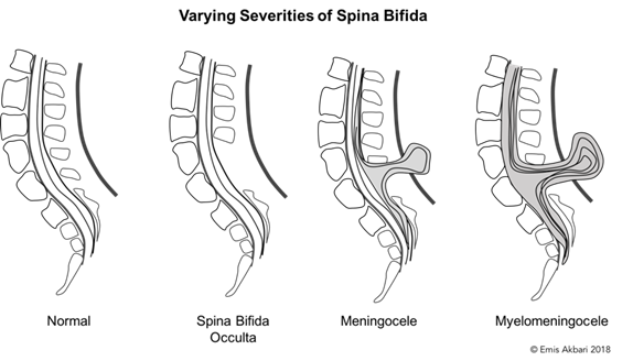
If the neural tube does not close properly, the results are referred to as neural tube defects. Spina Bifida, which can vary greatly in severity (see image), is the most common of neural tube defects. Prenatal folic acid supplements help prevent Spina Bifida. By the 20th day after conception, swellings at the top of the neural tube appear and form three structures: the hindbrain (rhombencephalon), the midbrain (mesencephalon), and the forebrain (prosencephalon). The hindbrain and the midbrain together comprise the brainstem. Further on in development, the hindbrain divides into the metencephalon (including the pons and the cerebellum), and the myelencephalon (medulla). The midbrain does not develop into further divisions; however, the forebrain further divides into the diencephalon and the telencephalon.
The table identifies the major divisions of the brain, their main associated structures and functions.
| Division | Structure | Function |
|---|---|---|
| Hindbrain | Medulla | Heart rate, breathing, blood pressure |
| Pons | Sleep – wake cycles | |
| Cerebellum | Balance, motor control | |
| Midbrain | Substantia Nigra | Motor control |
| Superior and Inferior Colliculi | Visual and auditory reflexes | |
| Forebrain | Cerebral Cortex | Cognition, motor and sensory processing. Highest level of processing. |
| Thalamus | Sensory processing, arousal | |
| Hypothalamus | Hormonal regulation, hunger, thirst, sexual and maternal behavior | |
| Hippocampus | Stress and memory | |
| Amygdala | Aggression, fear | |
| Corpus Callosum | Connection between the two hemispheres |
Communication in the central nervous system arises from nerve cells called neurons. Most of our neurons are produced prior to birth with peak production of 250,000 neurons per minute occurring around mid-pregnancy (Purves, Augustine, & Fitzpatrick, 2004). Neurons are brain cells that store and transfer information. As they are produced, they gradually move (migrate) to their permanent locations. Many of these early brain cells die prior to birth as the brain proliferates and prunes (Stiles & Jernigan, 2010).
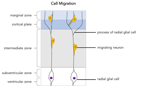
The generation of new cells is called cell proliferation. These new cells eventually develop into either neurons or glia (support cells within the central nervous system) and are produced within the ventricular zone. This area is adjacent to the brain’s ventricles and is the location of neuronal generation. The ventricular zone produces progenitor cells, which eventually develop into either neurons or glia. Radial glial cells are also produced here, and support the migration of new cells to their final destination. This movement of newly formed cells to their final destination is known as cell migration. This occurs when cells glide along the radial glial cells that emanate from the ventricular zone (see image below).
During cell proliferation, the embryo is very vulnerable to even slight environmental disturbances. One example of a resulting problem is microencephaly. This is a group of disorders characterized by a small brain due to errors in neural cell proliferation. Microencephaly can be caused when the embryo is exposed to toxins such as radiation, rubella, alcohol, or the zika virus.
Newly generated cells then begin to differentiate. Differentiation occurs when these newly formed cells acquire the characteristics of different types of neurons. Neurons form complex networks of fibers called dendrites and axons. Dendrites are fibers that radiate from the soma (cell body) and receive electrochemical information from other neurons. Each neuron has numerous dendrites. Each neuron also has one axon that extends away from the soma and transmits electrochemical information to the dendrites of another neuron.
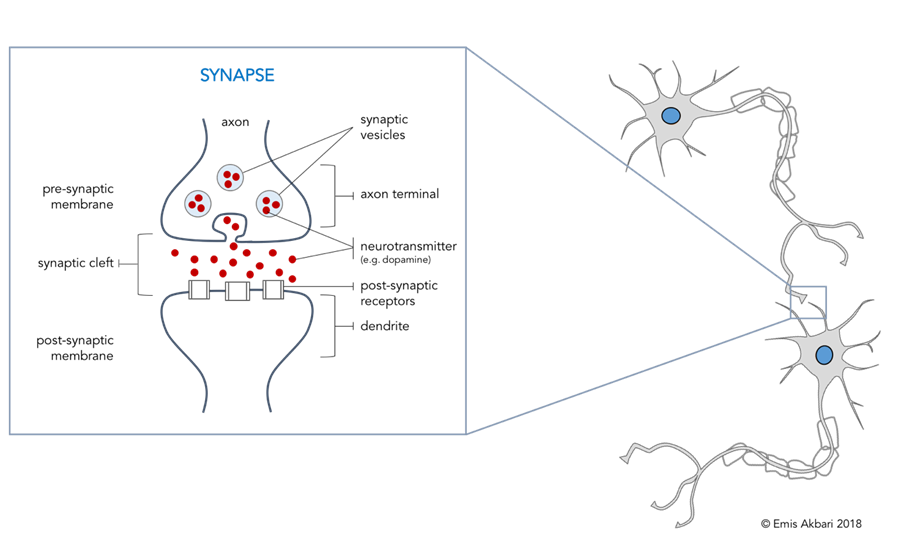
As neurons move to their final destinations, their axons start to extend in order to connect with other neurons (axonal outgrowth). The location of where one neuron’s axon (axon terminals) meets the dendrites of another neuron is known as a synapse. Synaptogenesis refers to the production of these synaptic connections. These tiny points of communication occur with the release of neurotransmitters, a chemical messenger of information. Neurotransmitters are stored in synaptic vesicles in the axon terminals (see image below).
Each neuron releases its own neurotransmitter with receptors on neighbouring neurons that are activated by the specific neurotransmitter that is released. An electrical signal originates in the presynaptic cell and is converted into a chemical signal (neurotransmitter) that can be transferred to the postsynaptic cell. By sending these chemical messages across the synapses, neurons communicate with each other. Scientists have identified over 60 neurotransmitters that are produced in presynaptic neurons and released into the synaptic gap to affect the postsynaptic neuron.
These synaptic connections get strengthened and the brain gets bigger and starts to look more like a mature brain with protrusions (gyrus) and grooves (sulcus) along the surface to accommodate the larger cortex.
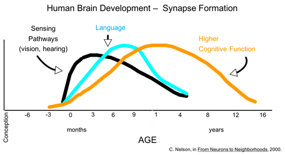
The first synapses are formed as early as the 23rd week of gestation, but most synapses develop later in the pregnancy and after birth, particularly during the first year of life. The timing of synapse formation varies from one area of the brain to another. For example, in the area of the brain responsible for vision (the occipital lobe) the greatest increases in synaptogenesis occur between 2 and 4 months of age. This contrasts with the middle frontal gyrus (believed to be involved in more advanced cognition), which has the peak number of synapses at 3 ½ years.
While synapses form and require space, approximately half of all brain cells die off. This programmed cell death is called apoptosis.
The brain overproduces synapses and then those that aren’t used are eliminated. This synaptic pruning occurs late in childhood and in adolescence. When synapses are pruned, the accompanying neurons become available for future development. Through childhood and adolescence, approximately 40% of synapses are pruned to arrive at the adult level of synapses. The timing of synaptic pruning varies depending on the area of the brain. The experiences an individual has have a major effect on both the overproduction and pruning of synapses of his/her brain. The image below demonstrates the developmental timing of synapse formation and subsequent pruning (Nelson, 2000).
Glial cells that support and feed the neurons (e.g., by myelination) continue to flourish throughout pregnancy and after birth. This causes substantial brain weight growth. Myelin is a fatty layer that coats axons and increases the efficiency of message transfer from one neuron to another. Glial cells are responsible for this myelination, (done by Oligodendrocytes in the central nervous system, and Schwaan cells in the peripheral nervous system), which starts to form in the last 3 months of pregnancy.
Myelinated axons are also referred to as white matter as opposed to grey matter, which is mostly made up of neuronal bodies, unmyelinated axons and glial cells. As neural fibers and myelination increase, the brain continues to increase in size. In fact, while most neurons have been myelinated by the baby’s second birthday, white matter continues to increase gradually and steadily through the 20’s. As with synaptogenesis, the process of myelination starts before birth but most of it occurs after the baby is born.
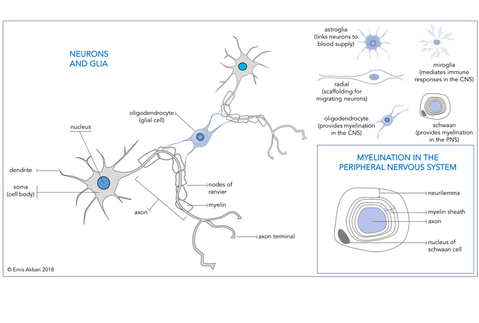
Infant nutrition plays an important role in the myelination of axons and breastfeeding is endorsed over formula feeding because infant formulas have been shown to be an inadequate source of the essential fatty acids important for myelination and healthy brain development (College of Family Physicians of Canada, 2004). See the table below for major events in the early prenatal period.
| Weeks post conception | What’s happening |
|---|---|
| 1 & 2 | Cells (referred to as a zygote) are multiplying and implanting in the uterine lining. |
| 3 & 4 | Now referred to as an embryo until week 9. Neural induction and neurulation occur. With the first wave of cell migration, early brain structures appear by the 20th day after conception. |
| 7 | Asymmetrical cells proliferation begins and continues until the middle of pregnancy. |
| 9 | Embryonic period ends; now referred to as a fetus. |
| 11 & beyond | Second wave of cell migration occurs; completed by the end of the 6th month. Cells become specialized for varying functions (differentiation) as neurons migrate to new locations and begin to form synapse (synaptogenesis). |
In summary, brain development starts at conception and continues through adolescence and beyond. The major brain developments that occur are: (1) neural induction and neurulation, then (2) cell proliferation and (3) cell migration, followed by (4) cell differentiation. Both (5) synaptogenesis and (6) myelination begin prenatally and continue for years after birth.
References
Belsky, J. & de Haan, M. (2011). Annual research review: Parenting and children’s brain development: The end of the beginning. Journal of Child Psychology and Psychiatry, 52(4), 409-428.
Berk, L. & Shanker, S. G. (2006). Child Development (Second Canadian Edition). Toronto: Pearson.
The Brain from Top to Bottom [Website]. Retrieved from http://thebrain.mcgill.ca/flash/index_d.html
College of Family Physicians of Canada (2004). Infant feeding policy statement 2004. Retrieved from: http://www.cfpc.ca/uploadedFiles/Resources/Resource_Items/Final_04Infant_Feeding_Policy_Statem ent.pdf
Founders’ Network. [Website]. Retrieved from http://www.founders.net/
Gilles, F. H., Green, B. E., Nelson, M. D., & Gwinn, J. L. (2012). The developing human brain: growth and adversities. Clinics in Developmental Medicine, 193(1), ALL-ALL.
Nelson, C. A. (2000). From neurons to neighborhood. The Science of Early Childhood Development. National Research Council Institute of Medicine.
Nelson, C. (2011). Neural development and lifelong plasticity. In D. Keating (Ed.), Nature and nurture in early child development (pp. 45-69). USA: Cambridge University Press.
Ralston, A. & Shaw, K. (2008). Gene expression regulates cell differentiation. Nature Education 1(1):127. Retrieved from http://www.nature.com/scitable/topicpage/gene-expression-regulates-cell-differentiation-931
Stiles, J., & Jernigan, T. (2010). The basics of brain development. Neuropsychology Review, 20(4), 327-348.

